Science
Behind the Condition

Causes of AVM

This is the heading

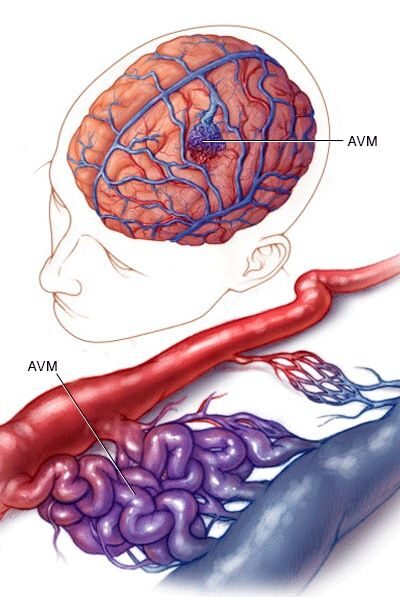
The exact cause of Arteriovenous Malformation (AVM) is not fully understood, but researchers believe it is primarily a result of abnormal blood vessel development during early stages of fetal growth. This condition arises when the arteries and veins form direct connections without the presence of capillaries, which typically regulate blood flow between them. While most AVMs are congenital (present at birth), they are not usually hereditary. Here are some key factors associated with the causes of AVM:

Congenital, Not Genetic: Although present at birth, AVMs are typically not inherited and do not run in families.

Abnormal Blood Vessel Formation: AVMs are thought to occur during embryonic or fetal development, leading to irregular connections between arteries and veins.

Rarely Linked to Environmental or External Factors: There is no strong evidence that lifestyle, environment, or other external factors play a role in causing AVM.

While the exact mechanisms remain unclear, ongoing research aims to uncover more details about how AVMs develop and the potential genetic or molecular pathways involved.

Types of AVM

Arteriovenous Malformations (AVMs) can occur in various parts of the body, each presenting unique challenges and symptoms. Understanding the different types helps in identifying and managing the condition effectively. The main types of AVM include:

Cerebral AVM: Found in the brain, this is the most common and well-known type. It can cause headaches, seizures, or even life-threatening bleeding (hemorrhage).

Spinal AVM: Occurs in or near the spinal cord, potentially leading to back pain, weakness, or paralysis if left untreated.

Pulmonary AVM: Affects the lungs, where abnormal connections between arteries and veins can lead to difficulty breathing, low oxygen levels, or stroke-like symptoms.

Peripheral AVM: Found in other areas of the body, such as arms, legs, or organs. These can cause pain, swelling, or skin changes.

Galenic AVM: A rare type that occurs near the vein of Galen in the brain, often detected in infants or young children.

Each type of AVM requires specific diagnosis and treatment approaches, emphasizing the importance of early detection and specialized care.

Pathophysiology of AVM

The pathophysiology of Arteriovenous Malformation (AVM) lies in the abnormal development of blood vessels, where arteries connect directly to veins without an intervening capillary network. This bypass disrupts normal blood flow and pressure regulation. Arteries, which carry high-pressure blood from the heart, directly transfer this pressure to veins, which are not designed to handle it. Over time, this can lead to vessel dilation, rupture, or bleeding.

In AVMs, the lack of capillaries impairs oxygen delivery to surrounding tissues, causing localized ischemia (oxygen deprivation). Additionally, the high blood flow through the AVM can "steal" blood from nearby normal tissues, exacerbating the problem. This abnormal vascular structure is thought to result from errors in angiogenesis during fetal development, but the exact molecular mechanisms remain unclear. Understanding this process is critical for developing effective treatments.

Risk Factors of AVM

The exact cause of Arteriovenous Malformations (AVMs) remains uncertain, and for most individuals, AVMs are congenital, meaning they are present at birth. However, several factors may increase the likelihood of having or developing AVM. These include:

Congenital Origins: AVMs are believed to form during fetal development, although they may not cause symptoms until later in life.

Genetic Syndromes: Rare genetic conditions, such as Hereditary Hemorrhagic Telangiectasia (HHT), can predispose individuals to AVMs.

Age and Gender: AVMs are often diagnosed in younger individuals, usually before the age of 40, and may be slightly more common in males.

While these factors are associated with AVMs, their presence does not guarantee the development of the condition. Many AVMs are discovered incidentally, highlighting the importance of awareness and medical evaluation for early diagnosis.

Symptoms of AVM

The symptoms of Arteriovenous Malformation (AVM) can vary widely depending on its location and size. While some AVMs remain asymptomatic and are discovered incidentally, others can cause significant health issues. Common symptoms include:

Neurological Symptoms: In brain AVMs, headaches, seizures, dizziness, or vision problems may occur.

Bleeding (Hemorrhage): A ruptured AVM can lead to sudden and severe symptoms, such as a stroke, due to bleeding in the brain or other affected areas.

Neurological Deficits: Spinal AVMs can result in back pain, weakness, or even paralysis if untreated

Neurological Deficits: Spinal AVMs can result in back pain, weakness, or even paralysis if untreated

Chronic Pain: Peripheral AVMs may cause localized pain, swelling, or skin discoloration.

Symptoms can range from mild to life-threatening, emphasizing the need for early diagnosis and treatment to prevent serious complications

Diagnosis of AVM

The diagnosis of Arteriovenous Malformation (AVM) typically involves a combination of clinical evaluation, imaging techniques, and sometimes invasive procedures. Doctors start by assessing symptoms, medical history, and physical examinations to identify potential signs of AVM. Advanced imaging is crucial for confirming the diagnosis and determining the AVM's location, size, and structure. Common diagnostic methods include:

Magnetic Resonance Imaging (MRI): Provides detailed images of the brain, spinal cord, or other affected areas, helping to detect AVMs and associated complications like bleeding.

Computed Tomography (CT) Scan: Useful in emergency settings to quickly identify bleeding or structural abnormalities.

Angiography: A specialized procedure using contrast dye and X-rays to map the blood vessels and visualize the AVM in detail.

Doppler Ultrasound: For peripheral AVMs, ultrasound can evaluate blood flow and vessel abnormalities.

Accurate diagnosis is essential for determining the most appropriate treatment plan and preventing further complications.